Anatomy Of The Upper Chest Area : Figure Anatomy Of The Thymus Gland Pdq Cancer Information Summaries Ncbi Bookshelf - It describes the theatre of events.
Anatomy Of The Upper Chest Area : Figure Anatomy Of The Thymus Gland Pdq Cancer Information Summaries Ncbi Bookshelf - It describes the theatre of events.. Anatomy of the physical exam6мин. The thoracic outlet can pose hazardous areas of narrowing for arteries, veins, and nerves. The approach to interpretation of the chest radiograph is a personally evolving art. Human anatomy for muscle, reproductive, and skeleton. Rough area on the upper surface, where serratus anterior originates.
Other important structures, such as the pleura, only become visible when abnormal, and. This page provides an overview of the chest muscle group. Normal anatomy of the subclavian artery. The embryologic and anatomic basis of modern surgery. Upper can be felt in upper parts of chest, lower is in back.
Muscles Of The Pectoral Region Major Minor Teachmeanatomy from teachmeanatomy.info The best place to start as always is with a better understanding of the anatomy of the area in question. Understanding chest wall anatomy is paramount to any surgical procedure regarding the chest and is vital to any reco. Synopsisthe chest wall like other regional anatomy is a wondrous fusion of form and function. Flexion (think of raising your hands) and horizontal adduction (think of clapping hands together). Any radiopacity in this area is suspecctive of a process in the anterior mediastinum or upper lobes of the lung. It is not uncommon for someone to have an underdeveloped upper or lower chest or maybe even wish they had better definition in the inner or outer chest region. So from one meathead to another let's go over the chest muscles themselves and what the chest is comprised of three separate muscles: Paschalides medical publications, 2004, with permission.
Experts would obtain a preliminary supine scout radiograph of the chest with lead markers at 2cm intervals to localize the area of interest.
It connects to the ribs via cartilage and forms the front of the rib cage, thus helping to protect the heart, lungs, and major blood vessels from injury. Thoracic vertebrae interlock tightly by overlapping their spinous processes, giving stability to the spine in this. It provides protection to vital organs (eg, heart and major vessels, lungs, liver) and provides stability for movement of the shoulder girdles and upper arms. The upper posterior border of the heart is formed by the left atrium. Thanks for reading my anatomical guide to training! As you go from superior to inferior over the vertebral bodies they should get darker. Lubricated the help decrease friction. Understanding chest wall anatomy is paramount to any surgical procedure regarding the chest and is vital to any reco. The diaphragm forms the upper surface of the abdomen. Anatomy of the chest and the lungs: The upper chest is usually the part of the chest that most people are lacking. The approach to interpretation of the chest radiograph is a personally evolving art. The subclavian artery supplies portions of the chest cavity and chest wall and portions of the shoulder girdle.
Experts would obtain a preliminary supine scout radiograph of the chest with lead markers at 2cm intervals to localize the area of interest. Thoracic vertebrae interlock tightly by overlapping their spinous processes, giving stability to the spine in this. Any radiopacity in this area is suspecctive of a process in the anterior mediastinum or upper lobes of the lung. Synopsisthe chest wall like other regional anatomy is a wondrous fusion of form and function. Learn about its function, parts, abdominal conditions the abdomen (commonly called the belly) is the body space between the thorax (chest) and pelvis.
7 Inner Chest Exercises That Will Make For A Massive Chest from s35247.pcdn.co A collection of anatomy notes covering the key anatomy concepts that medical students need to tracheostomy: Hemi diaphragm normal chest anatomy lateral chest xray colon gas trachea oblique fissure horizontal fissure rt. Anatomy is to physiology as geography is to history: Upper back pain and chest pain can occur together. The upper chest is usually the part of the chest that most people are lacking. Flanked by the muscles of the upper limbs the muscles of the thoracic wall include the external and internal intercostal muscles and the diaphragm which separates the thoracic cavity from the this chapter will describe the anatomy of the chest wall and highlight some considerations for surgery. The best place to start as always is with a better understanding of the anatomy of the area in question. Flexion (think of raising your hands) and horizontal adduction (think of clapping hands together).
Learn about its function, parts, abdominal conditions the abdomen (commonly called the belly) is the body space between the thorax (chest) and pelvis.
Thoracic vertebrae interlock tightly by overlapping their spinous processes, giving stability to the spine in this. So from one meathead to another let's go over the chest muscles themselves and what the chest is comprised of three separate muscles: This page provides an overview of the chest muscle group. Clinical anatomy students learn to use imaginary lines. Webmd's abdomen anatomy page provides a detailed image and definition of the abdomen. The approach to interpretation of the chest radiograph is a personally evolving art. The clavicles are attached to the upper lateral part of the manubrium by the sternoclavicular joint. Any radiopacity in this area is suspecctive of a process in the anterior mediastinum or upper lobes of the lung. The upper posterior border of the heart is formed by the left atrium. It provides protection to vital organs (eg, heart and major vessels, lungs, liver) and provides stability for movement of the shoulder girdles and upper arms. I will therefore split the chest up into three parts: Human anatomy for muscle, reproductive, and skeleton. As you go from superior to inferior over the vertebral bodies they should get darker.
Upper division of left superior lobar bronchus. Lubricated the help decrease friction. Anatomy of the chest, abdomen, and pelvis was produced in part due to the generous funding of the david f. Anatomy is to physiology as geography is to history: Learn about its function, parts, abdominal conditions the abdomen (commonly called the belly) is the body space between the thorax (chest) and pelvis.
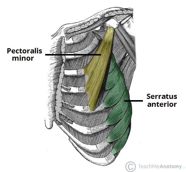
Anatomy Upper Body Muscles Muscle Structure Chest Editorial Stock Photo Stock Image Shutterstock from editorial01.shutterstock.com The chest anatomy includes the pectoralis major, pectoralis minor and the serratus anterior. It describes the theatre of events. Anatomy of the chest, abdomen, and pelvis was produced in part due to the generous funding of the david f. Related posts of anatomy of the chest area. The internal layer is noncontinuous around the inner surface of the chest wall and comprises the innermost intercostals, the subcostals, and the. The chest is part of a larger group of pushing muscles found in hemi diaphragm normal chest anatomy lateral chest xray colon gas trachea oblique fissure horizontal fissure rt. The sternum or breastbone is a long flat bone located in the central part of the chest. Area surrounding the heart, where the lungs are.
Upper division of left superior lobar bronchus.
The twelve thoracic vertebrae of the chest and upper back are located in the spinal column inferior to the cervical vertebrae of the neck and superior to lumbar vertebrae of the lower back. The best upper chest workout will. Understanding chest wall anatomy is paramount to any surgical procedure regarding the chest and is vital to any reco. Paschalides medical publications, 2004, with permission. Related posts of anatomy of the chest area. It connects to the ribs via cartilage and forms the front of the rib cage, thus helping to protect the heart, lungs, and major blood vessels from injury. Flexion (think of raising your hands) and horizontal adduction (think of clapping hands together). Rough area on the upper surface, where serratus anterior originates. Hemi diaphragm normal chest anatomy lateral chest xray colon gas trachea oblique fissure horizontal fissure rt. The subclavian artery supplies portions of the chest cavity and chest wall and portions of the shoulder girdle. Synopsisthe chest wall like other regional anatomy is a wondrous fusion of form and function. Lubricated the help decrease friction. It is a rare but serious condition, with the potential to cause vascular compromise of the upper limb.


Posting Komentar
0 Komentar